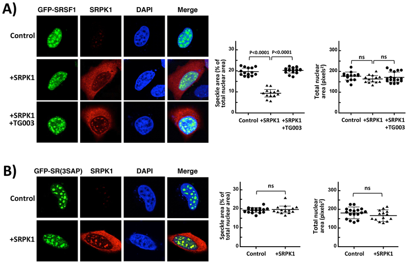
Figure 2: SRPK1 Over-expression Influences SRSF1 Subnuclear Localization.
A) Confocal microscopy of HeLa cells expressing GFP-SRSF1 with and without co-expression of SRPK1. B) Confocal microscopy of GFP-SR(3SAP) in the absence and presence of SRPK1 expression. All speckle areas and total nuclear areas for cells (n=13–16 in (A) and (B)) were calculated from replicate experiments using Image J and displayed as a dot plot with mean values ± S.D. All P-values were calculated using a one-way ANOVA test. P-values above 0.05 are considered nonsignificant (ns). Control reflects cells over-expressing GFP-SRSF1 or GFP-SR(3SAP) but not transfected with the SRPK1 vector.

