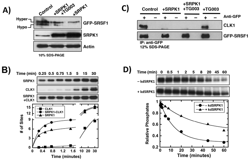
Figure 3: Regulating SRSF1 Phosphorylation Through The SRPK1-CLK1 Complex.
A) Effects of SRPK1 over-expression on GFP-SRSF1 phosphorylation state in HeLa cell extracts in the absence and presence of TG003. GFP-SRSF1, SRPK1 and actin were detected using anti-GFP, SRPK1 and actin antibodies. B) Single-turnover kinetic assay for SRSF1 phosphorylation by SRPK1, CLK1 and the SRPK1-CLK1 complex. Y-axis data are normalized to total SRSF1 concentration to obtain # of sites phosphorylated. SRPK1=CLK1= 1 μM, SRSF1= 100 nM, ATP= 50 μM. Data fits are presented in Supplementary Table S1. C) Co-immunoprecipitation of GFP-SRSF1 and endogenous CLK1 in HeLa cell extracts in the absence and presence of SRPK1 over-expression. GFP-SRSF1 and CLK1 were detected using anti-GFP and CLK1 antibodies. D) PP1-dependent dephosphorylation of CLK1-phosphorylated SRSF1 in absence and presence of kdSRPK1. CLK1=PP1=kdSRPK1= 2 μM, SRSF1= 200 nM, TG003= 50 μM, ATP= 100 μM. Data fits are presented in Supplementary Table S2. Controls reflect cells over-expressing GFP-SRSF1 but not transfected with the SRPK1 vector.

