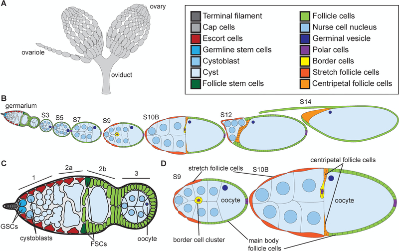
Figure 1:
Overview of Drosophila oogenesis. (A) Schematic of ovaries and oviduct. (B) Schematic of ovariole with the stages of development indicated. (C) Schematic of germarium. (D) Schematic of S9 and S10B follicles. Adult female fly has two ovaries that are comprised of ~15 ovarioles, or chains of sequentially maturing follicles (A). Follicle development is separated into 14 morphological stages (B), from the germarium to S14. The germarium is at the anterior tip of the ovariole (B), is broken into 4 regions (1, 2a, 2b, and 3; C) and contains both germline (bright cyan) and somatic or follicle stem cells (dark green). Two to three germline stem cells (GSCs, bright cyan) reside at the anterior of region 1 in a niche comprised of the terminal filament (dark gray), cap (light gray), and anterior escort (red) cells (C). The GSCs divide asymmetrically to self-renew and generate a cystoblast (C, light cyan). The cystoblast goes on to have four incomplete and synchronous cell divisions to generate a 16-cell cyst (light blue). In region 3, the cyst will differentiate into 15 nurse cells (nuclei are light blue) and one oocyte (nucleus is dark blue) (C). At the 2a/2b boundary in the germarium (C), the follicle stems cells (FSCs, dark green) reside and give rise to all of the somatic, follicle cells (light green) that will encase the 16-cell cysts. These follicle cells will subsequently differentiate into a number of subtypes. By S9 (D), four different types of follicle cells are observed: the purple polar cells which specify the poles, the yellow migrating border cell cluster (comprised of the anterior purple polar cells and surrounding yellow border cells), the squamous, dark orange stretch follicle cells, and the green main body follicle cells. By S10B (D), another follicle cell subtype is observed, the centripetal follicle cells in light orange. An additional type of follicle cells, stalk cells, are not shown but connect the follicles to each other.

