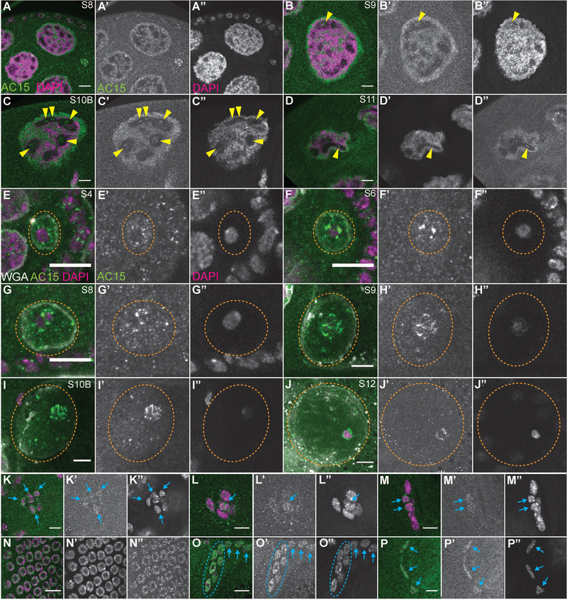
Figure 10:
AC15 nuclear actin localizes to the DNA. (A-P”) Zoomed in images of different cell types from maximum projections of 2–4 slices of confocal stacks of the indicated stages of wild-type (yw) follicles. (A-D, K-P) Magenta = DAPI, Green = AC15. (E-J) Magenta = DAPI, Green = AC15, and White = Wheat germ agglutinin (WGA, marks nuclear envelope). (A’-P’) White = AC15. (A”-P”) White = DAPI. (A-D”) Nurse cells. (E-J”) Oocytes. (K-M”) Border cells. (N-N”) Main body follicle cells. (O-O”) Centripetal follicle cells. (P-P”) Stretch follicle cells. Within the nurse cells, AC15 largely colocalizes with the DNA (A-D”), however it also labels puncta within regions devoid of chromatin in S9–11 (yellow arrowheads). In the oocyte (nuclei circled by orange dashed line), AC15 nuclear actin structure changes with development (E-J). In S4 oocytes, AC15 nuclear actin exhibits a speckled appearance throughout the nucleoplasm, with bright puncta adjacent around the edge of the chromatin (E-E”). This enrichment of AC15 puncta around the DNA in the oocyte increases in S4–8 (F-G”), and then appears to form filaments that encircle the chromatin in S9–11 (H-I”). In S12 oocytes, AC15 nuclear actin becomes more diffuse, but still surrounds the DNA (J-J”). AC15 nuclear actin is also observed in the somatic cells. In the migrating border cells, AC15 nuclear actin is weakly observed in all of the cells (both border and polar cells) in the early stage of migration (K-K”, blue arrows). Towards the end of the border cell migration (late S9 and S10A), AC15 nuclear actin appears restricted to a subset of the cells (L-M”, blue arrows); given the localization of the AC15 positive nuclei, we hypothesize that they are the polar cells. AC15 nuclear actin is observed in all of the other follicle cell populations, including the main body follicle cells (N-N” and blue arrows in O-O’), centripetal follicle cells (circled in blue dashed line), and the stretch follicle cells (P-P”, blue arrows). Scale bars = 10μm.

