Figure 5:
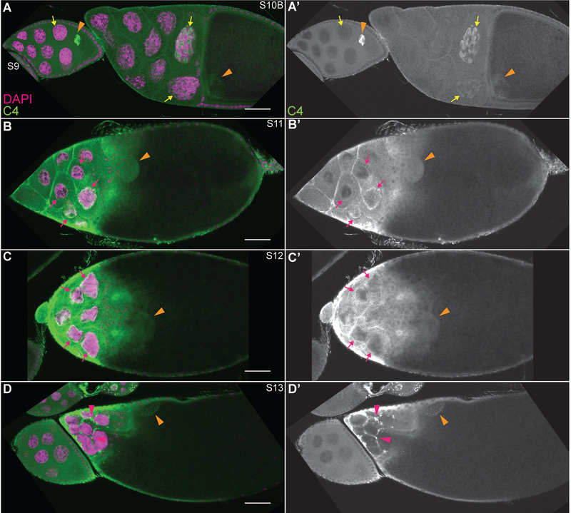
C4 nuclear actin is significantly reduced and relocalizes during late oogenesis. (A-D”) Maximum projections of 2–4 slices of confocal stacks of the indicated stages of wild-type (yw) follicles. (A-D) Magenta = DAPI, Green = C4. (A’-D’) White = C4. During the late stages of follicle development, C4 nuclear actin goes from a tubular structure observed at a low frequency in S10B (A-A’, yellow arrows), to localizing to a ring just underneath the nuclear envelope in S11–12 (B-C’, magenta arrows). Finally, during S13, C4 labels puncta that appear to be in either the cytoplasm of the nurse cells or the stretch follicle cells enveloping them (D-D’, magenta arrowheads). C4 nuclear actin is observed throughout the nucleoplasm of the oocyte (A-D’, orange arrowheads). Note that not all cells with C4 nuclear actin are marked; marks are used to point out specific cells in which the C4 nuclear actin is readily apparent in the focal planes shown. Scale bars = 50μm.

