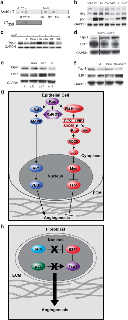
Figure 7.
Regulation of Tsp-1 expression in fibroblasts. (a) Schematic depiction of the domain structure of the SV40 LTC (LT) protein and the various mutants: LTK1-point mutation in the Rb binding motif, LT350-deletion mutation that only contains the first 350 amino acids of LT, LTD402H- point mutation in the p53 binding motif, J*-point mutation in the J domain and Cul7*-deletion mutation in the Cul7 binding motif; Immunoblot analysis of (b) Tsp-1, Myc, p53, and GAPDH expression in immortalized human dermal fibroblasts expressing the Large T mutants depicted in Figure 4a. (c) Tsp-1 and GAPDH expression in human dermal fibroblasts expressing hTERT (−) alone or in combination with: SV40 LTC (LT), shp53 (shp53), LTD402H (402), shp53 and LTD402H, LT350 (350), and shp53 and LT350. (d) Tsp-1, E2F1 and GAPDH expression in human dermal fibroblasts expressing hTERT alone (−) or in combination with shRNA specific for E2F1 (shE2F1a and shE2F1b). (e) Tsp-1, E2F1 and GAPDH expression in human dermal fibroblasts expressing hTERT alone (−), or in combination with pCMV, pCMVE2F1 (E2F1) or Large T (LT), numbers represent relative intensity of Tsp-1 in each lane normalized to GAPDH. (f) Tsp-1, E2F1 and GAPDH expression in by human dermal fibroblasts expressing hTERT alone (−), or in combination with SV40 LTC (LT), shp53, and shp53 plus pCMVE2F1 (E2F1). (g) Schematic diagram of Tsp-1 regulation in epithelial cells. (h) Schematic diagram of Tsp-1 regulation in fibroblasts. Black lines in western blots represent cases where lanes were digitally removed from the scanned image.

