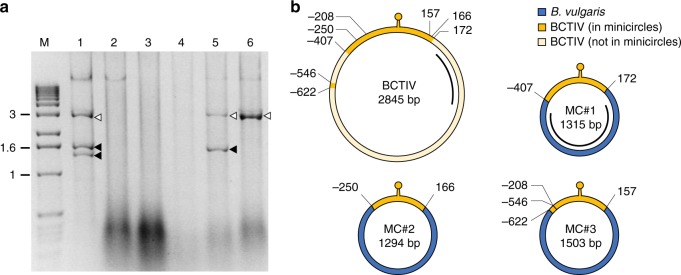
Fig. 1.
Identification of chimeric minicircles in field-grown sugar beet plants. a DNA fragments obtained after RCA and EcoRI digestion of DNA samples from different plants (lanes 1–6) collected from a field in Iran. The putative full-length BCTIV genome and smaller molecules are indicated by white and black arrows, respectively. Lane M, molecular markers with the size (kb) indicated on the left. Source data are provided as Supplementary Data 5. b Schematic representation of BCTIV and the three minicircles obtained from some of the BCTIV-infected field samples shown in a; MC#1 originates from plant 5, MC#2 and #3 from plant 1. The BCTIV and the minicircle genomes are represented in circular form; yellow and blue represent the BCTIV and the B. vulgaris genome-derived elements, respectively. The recombination coordinates on the BCTIV genome and the minicircles are indicated in nucleotides in relation to the conventional start of the BCTIV genome located at the stem-loop region (marked as a yellow lollipop). The locations of the MC#1- and BCTIV-specific probes used for subsequent southern blot analysis are represented by black lines. The complete minicircle sequences are reported in Supplementary Data 2

