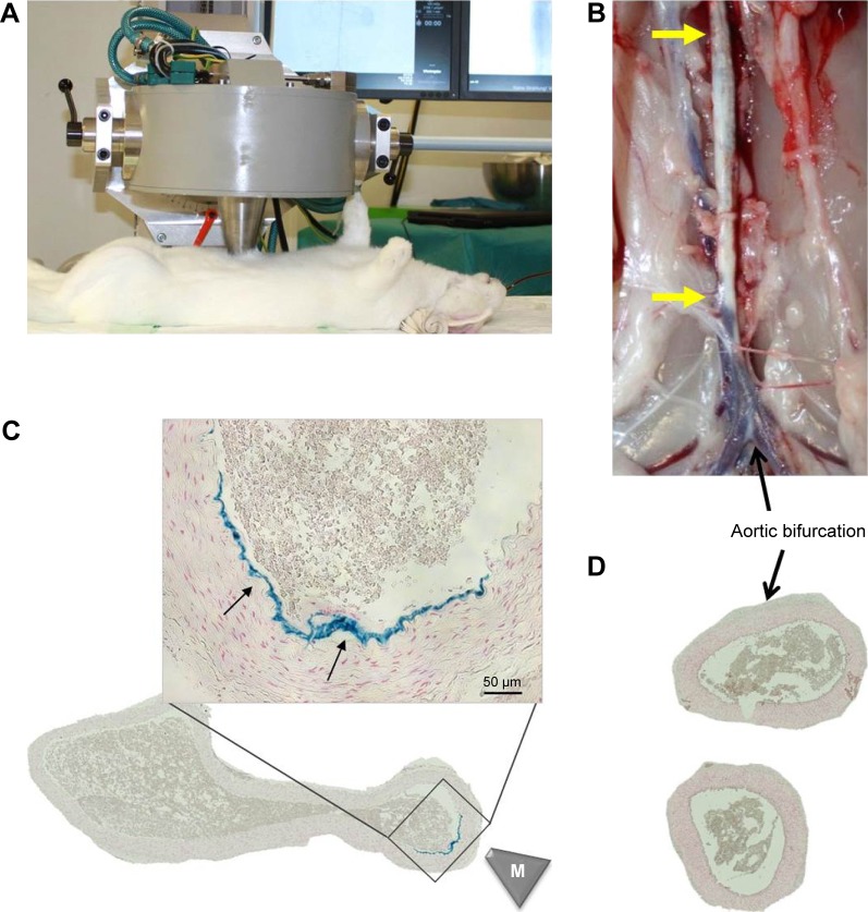
Figure 4.
Magnetic targeting setup and efficacy.
Notes: SPIONs were magnetically targeted to the injured aortic region directly after the ballooning. (A) Experimental setup and placement of magnetic tip are shown. (B) Following the magnet exposure for 30 minutes, the animals were sacrificed and the excised aorta analyzed histologically. Atherosclerotic plaque is visible as indicated by yellow arrows. (C) The presence of iron in the targeted region was visualized by Prussian blue staining (arrows). Scale bar: 50 µm. (D) No iron accumulation was detected in aortic bifurcation region by histology. The overview images of the artery cross-sections were taken at ×10 objective magnification. M indicates the tip of the magnet.
Abbreviations: SPIONs, superparamagnetic iron oxide nanoparticles.

