Figure 1.
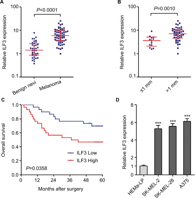
ILF3 is increased in melanoma and correlated with poor prognosis.
Notes: (A) ILF3 expression levels in 37 benign nevi and 60 primary melanoma tissues were determined by qRT-PCR. Results are presented as median with IQR; P<0.0001 by Mann–Whitney U-test. (B) ILF3 expression levels in melanoma tissues categorized based on tumor thickness; n=13 for ≤1 mm thick group, n=47 for >1 mm thick group; P=0.0010 by Mann–Whitney U-test. (C) Kaplan–Meier survival analysis of the correlation between ILF3 expression levels and overall survival of these 60 melanoma patients. ILF3 median expression level was used as the cutoff. P=0.0358 by log-rank test. (D) ILF3 expression levels in human epidermal melanocytes HEMa-LP and melanoma cell lines SK-MEL-2, SK-MEL-28, and A375 were determined by qRT-PCR. Results are presented as mean ± SD based on three independent experiments. ***P<0.001 by one-way ANOVA.
Abbreviation: qRT-PCR, quantitative real-time PCR.

