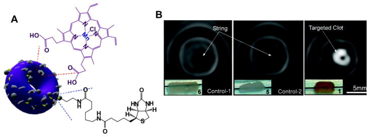
Figure 4.
Self-assembled nanobialys. A) The structure of Mn(III) porphyrin nanobialys. Mn(III) porphyrin provided MRI contrast. B) MRI of fibrin-targeted nanobialys (1) (right) or control group with biotin and no metal (6)(left), or no biotin with metal (5)(middle) bound to cylindrical plasma clots measured at 3.0 T. Permission obtained from Ref.[70]. Copyright (2008) by the American Chemical Society.

