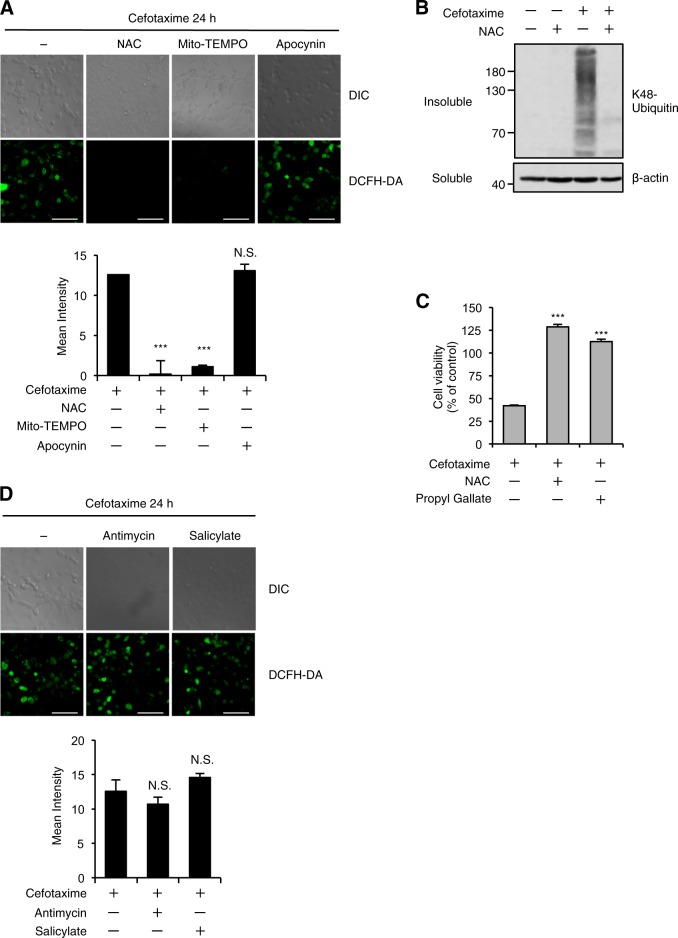
Fig. 2. Oxidative stress is responsible for the ALIS formation and cell death induced by the cephalosporins.
a, d HT1080 cells were treated with the indicated reagents for 24 h, and then treated with 10 μM DCFH-DA. Fluorescence images (upper panel) and intensity (lower panel) of HT1080 cells were acquired as described in the materials and methods section. Cell morphology was determined by Nomarski differential interference contrast (DIC) microscopy. Data shown are the mean ± SD. Significant differences were determined by one-way ANOVA, followed by Tukey–Kramer test; ***p < 0.001; N.S., not significant. Scale bar, 50 μm. b HT1080 cells were treated with the indicated reagents for 36 h, and then the detergent-soluble and -insoluble fractions were subjected to immunoblotting with the indicated antibodies. c HT1080 cells were treated with the indicated reagents for 48 h, and subjected to cell viability assay. Data shown are the mean ± SD. Significant differences were determined by one-way ANOVA, followed by Tukey–Kramer test; ***p < 0.001. Cefotaxime (1 mg/ml), NAC (1 mM), Mito-TEMPO (10 µM), Apocynin (100 µM), Propyl Gallate (20 µM), Antimycin (10 ng/ml), Salicylate (10 mM)

