Fig. 4.
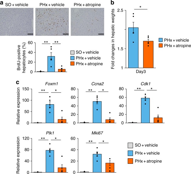
Muscarinic signals are involved in acute liver regenerative responses after PHx. a (Upper panels) Representative images of liver sections immunostained for BrdU on day 2 after sham operation for PHx (SO) and PHx in vehicle-treated mice and after PHx in atropine-treated mice. Scale bars indicate 100 µm. (Lower panel) BrdU-positive hepatocyte ratios in SO + vehicle mice (n = 4), PHx + vehicle mice (n = 5), and PHx + atropine mice (n = 5). b Fold changes in hepatic weights 3 days after surgery in PHx + vehicle mice (n = 5) and PHx + atropine mice (n = 5). Hepatic weights were divided by those obtained immediately after surgery. c Relative expressions of Foxm1, its target genes, and MKi67 in the liver 2 days after SO (n = 4) and PHx (n = 5) in vehicle-treated mice and PHx (n = 5) in atropine-treated mice. *P < 0.05; **P < 0.01 assessed by one-way ANOVA followed by Bonferroni’s post hoc test (a and c) or assessed by unpaired t test (b). n.s., not significant

