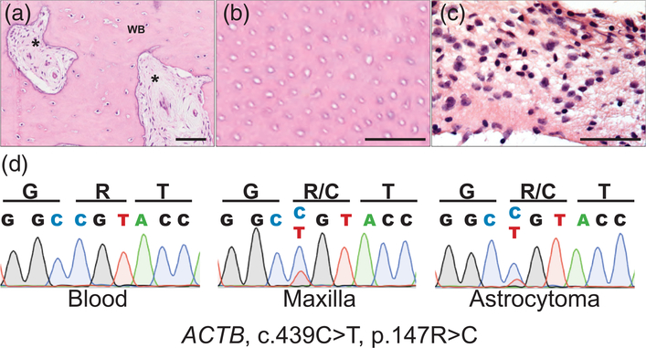
FIGURE 2.
ACTB mutation in maxillary lesion and pilocytic astrocytoma. (a) Representative 20× histology from an area of the maxillary lesion, which included areas of fibrosis (asterisk) within areas of woven bone (WB). (b) 40× view of tubular-like structures consistent with dentinal tubules. (c) 40× histology of smear of brain stem lesion demonstrates monomorphic population of glial tumor cells with elongated pilocytic processes in myxoid matrix, without Rosenthal fibers or eosinophilic granular bodies. (d) Sanger sequencing of ACTB demonstrates multilineage somatic c.439C>T, p.147R>C mutation, present in both the maxillary bone lesion (middle) and astrocytoma (right), which is absent in blood (left) [Color figure can be viewed at wileyonlinelibrary.com]

