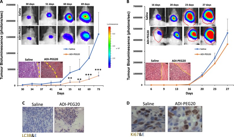
Fig. 2. In vivo assessment of ADI-PEG20 in mice bearing ASS1 negative and positive GBM tumour.
a, b CD−1 nude mice were stereotactically injected with LN229/ GFP-Luc or U87/ GFP-Luc cells as described in methods. On day 7 and 9 post injection of cells (first detection of luciferase activity by BLI for LN229/GFP-Luc and U87-GFP/Luc cells respectively) animals were injected IM with 0.9% saline or 5IU ADI-PEG20. Treatments were repeated on a weekly basis for the duration of the study and tumour growth assessed by BLI analysis twice per week. Bioluminescent images of representative animals are shown starting from day 30 onwards for animals bearing LN229 tumours and from day 16 onwards for mice bearing U87 tumours. Colour depicts relative luciferase signal. Bioluminescence signal plotted as photons/sec against time in days. P values: **P < 0.01, ***P < 0.001. The error bars are +/− 1 SD from seven animals. a, b Inset: H&E staining of representative tumour sections (x 1.25). T tumour, NB normal brain. c, d IHC analysis for LC3B and Ki67 in tumour sections from ADI-PEG20 and saline treated animals

