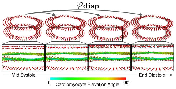
Fig. 2.
Displacement-based registration. The deformation mapping φdisp at each red point above and below the cDTI slice measures the motion between the cDTI acquisition phase (e.g., MS) and the target cardiac phase (e.g., ED). The corresponding deformation gradient F(eq. 1) is used to register the cardiomyocyte orientation vectors e⃗1 (eq. 2).

