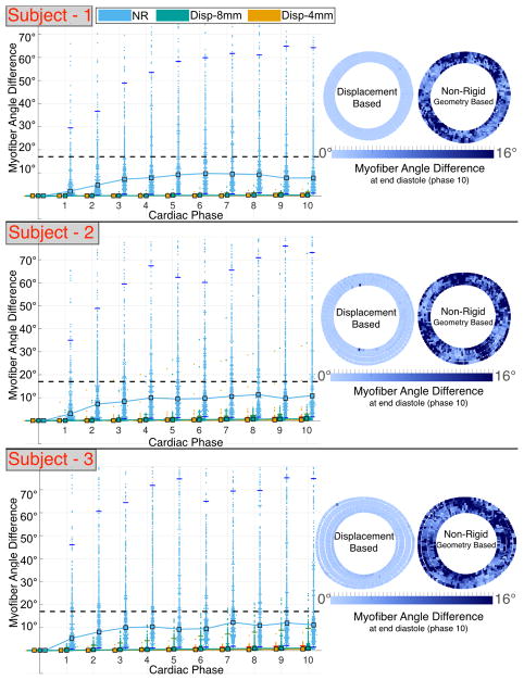
Fig. 4.
Angular differences between ground truth and registered (MS mapped to ED) cardiomyocyte orientation using displacement-based and NR approaches in subject-specific computational phantoms based on N = 3 volunteers. Left: median (square markers) and 95% CI (horizontal tick marks). The dashed line corresponds to the CODE cDTI cone of uncertainty [14]. Right: pointwise registration error at ED.

