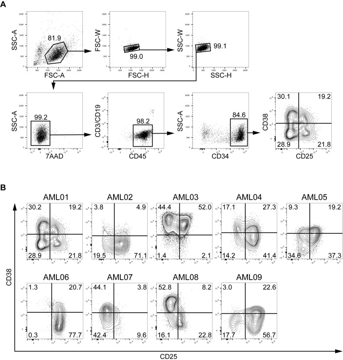
Fig 1. Gating strategy and flow-cytometric analysis of AML cells.
A, Cells from AML01 were analyzed by flow cytometry. After discrimination of doublet and dead cells, the cells were gated as Lin–CD45low cells, and then CD34+ cells were analyzed for CD38 and CD25 expression. B, CD38 and CD25 expressions on Lin–CD45lowCD34+ population in nine AML patients are shown as contour plots. Percentages of each cell fraction are indicated in the plot areas.

