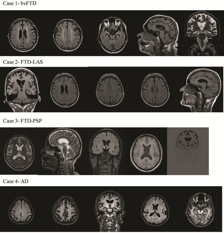
Fig 2. Brain MRI (T1, T2 and Flair) of C9orf72 expansions in the Bulgarian cohort (Case 1, 2, 3, 4).
Case 1—Bilateral frontal and temporal atrophy with very mild asymmetry that was more pronounced in the left hemisphere and hippocampal atrophy; Case 2—Mild frontal atrophy with very mild asymmetry that was more pronounced in the left frontal lobe and very mild hippocampal atrophy; Case 3—Mild hippocampal atrophy; Case 4—Generalized cortical atrophy, including posterior atrophy and bilateral hippocampal atrophy.

