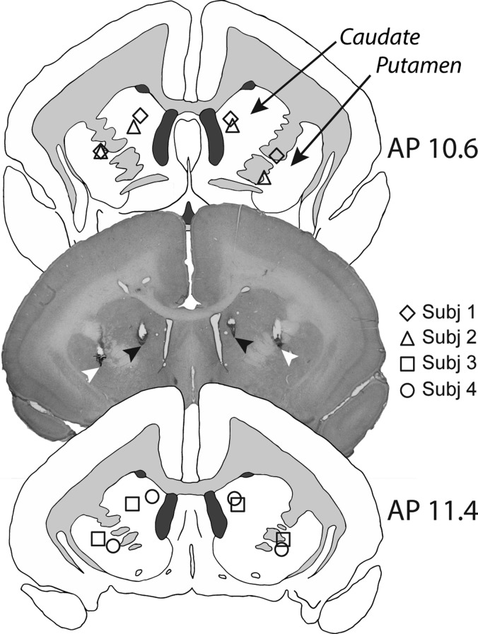
Figure 2.
Schematics showing the intra-cerebral cannulae placements in the medial caudate and putamen for each subject, in AP planes 10.6 and 11.4. In the representative histological section (taken from Subject 3), black arrows show the placement of the tip of the infusion cannulae in the medial caudate, and white arrows the placement of the tip of the infusion cannulae in the anterior putamen.

