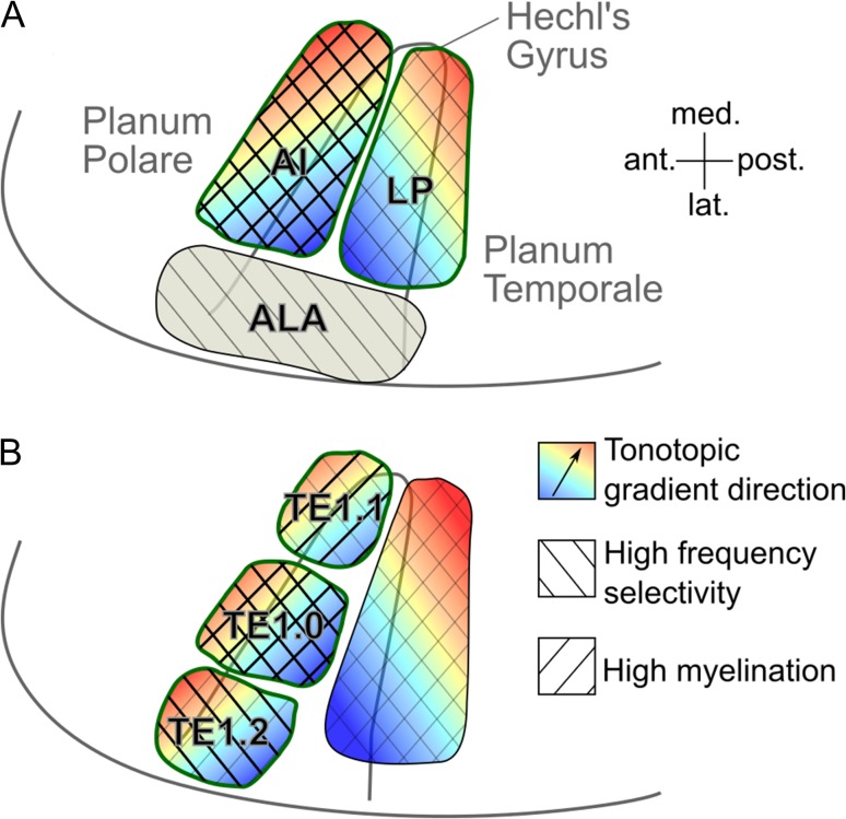
Figure 8.
Plausible models of human auditory cortex organization based on the current data and previous postmortem anatomical results. (A) Likely model under the assumption that the human core contains both tonotopic gradients on HG, and only occupies the medial and central parts of the gyrus. The green outlines designate core areas. The background hatch indicates increased frequency selectivity and/or myelin content (the degree of increase is indicated by the hatch weight). The area labels are in accordance with Rivier and Clarke (1997) and Wallace et al. (2002): AI = primary auditory area; LP = lateroposterior area; ALA = anterolateral area. (B) Likely model under the assumption that the human core only contains the anterior tonotopic gradient on HG, and occupies the gyrus’s full length. The area labels are in accordance with Morosan et al. (2001).

