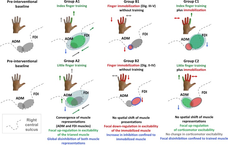
Figure 7.
Synopsis of within-area reorganization in right M1HAND observed in groups A1, B1, C1 and A2, B2 and C2. The left panels illustrate the preinterventional state with the gray areas reflecting the cortical representations of the FDI and ADM muscle in the right M1HAND. The arrows close to the schematic drawings of the hand summarize changes in learning performances for the trained and nontrained fingers. The gray shading illustrates “absence of intervention,” the green shading illustrates “training,” and the red shading illustrates “immobilization.” The arrows close to the schematic drawing of the central sulcus illustrate the direction of intervention-specific changes in muscle representations and intracortical inhibition.

