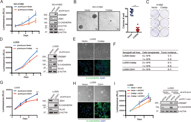
Figure 5. CREBBP re-expression in CREBBP-deleted human SCLC cells inhibits transformation.
A, CellTiter-Glo viability assay of ectopic expression of CREBBP in human CREBBP-deficient NCI-H1882 cells (n= 3 independent experiments). *, p<0.05. Immunoblotting of CREBBP, ZEB1, ASCL1 and E-CADHERIN were performed in these cells.
B, Anchorage-independent assay to test impact of CREBBP overexpression on growth of NCI-H1882 cells in soft agar. Cells were seeded at 1.0×10⁵ cells/well (6 well plate). n = 3 independent experiments. ***, p<0.001.
C, Colony formation assay to test impact of CREBBP overexpression on ability of NCI-H1882 cells to grow when plated at low density (n= 3 independent experiments). 3 representative technical replicates per condition were shown.
D, CellTiter-Glo viability assay of CREBBP overexpression in human CREBBP-deficient LU505 cells (derived from a PDX tumor). n= 3 independent experiments. ***, p<0.001. Immunoblotting of CREBBP, ZEB1, E-CADHERIN and SLUG were performed in these cells.
E, Representative phase contrast microscopic photos of LU505 cells with or without CREBBP overexpression (Upper). Scale bar = 100μm. Representative immunofluorescence images of E-CADHERIN staining in LU505 cells with or without CREBBP overexpression (Lower). DAPI was used as a nuclear stain. Original magnification, 40×.
F, Summary of differences in tumor-initiating ability of LU505-vector, LU505-CREBBP and LU505-CDH1 cells upon transplantation of 5 × 10⁶ cells or 1 × 10⁶ cells into immunocompromised NSG mice.
G, CellTiter-Glo viability assay of CDH1 overexpression in LU505 cells (n= 3 independent experiments). ***, p<0.001. Immunoblotting of ZEB1, E-CADHERIN and SLUG were performed in these cells.
H, Representative phase contrast microscopic photos of LU505 cells with or without CDH1 overexpression (Upper). Scale bar = 100μm. Representative immunofluorescence images of E-CADHERIN staining in LU505 cells with or without CDH1 overexpression. DAPI was used to stain nuclear. Original magnification, 40×.
I, CellTiter-Glo viability assay of shRNA-mediated CDH1 knockdown on the proliferation suppression induced by CREBBP overexpression in LU505 cells (n= 3 independent experiments). *, p<0.05. Re-expression of CREBBP and knockdown of CDH1 in this cell line were validated by immunoblotting.

