
Figure 3.
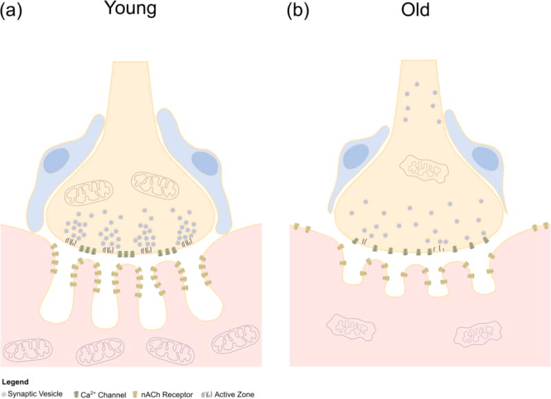
Cellular and molecular changes in the aged NMJ. (a) The young NMJ is characterized by deep junctional folds along the postsynapse containing high concentrations of nAChRs at the peaks. Active zones are organized on the motor axon terminal to lie in direct apposition to the junctional folds of the postsynapse. Vesicles containing acetylcholine aggregate at active zones where they are available to release acetylcholine in close proximity to nAChRs. Mitochondria are abundant throughout the pre- and postsynapse to support the high energy demands of cholinergic transmission. (b) With aging, the junctional folds of the postsynapse become shallow, while nAChRs are less concentrated along the peaks of the folds. In addition to postsynapses, nAChRs are found in extra-synaptic areas of the muscle fiber membrane. Mitochondria are damaged and fewer in the postsynaptic region of the muscle fiber. Active zones are lost in the motor axon terminal. Aggregation of vesicles containing acetylcholine near the terminal membrane is lost and vesicles are present away from the terminal along the axon. Dysfunctional megamitochondria are present in the motor axon terminal. Ensheathment of the NMJ by perisynaptic Schwann cells is lost as they migrate away from the motor axon terminal.