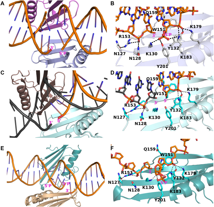
Figure 1.
Structures of ARTD2WGR bound to different DNA breaks. (A) Cartoon representation of ARTD2WGR bound to DNA breaks with 5′-phosphate. Two protein monomers (light blue and pink) and four DNA molecules, representing two double-stranded DNAs were present in the asymmetric unit. (B) Zoomed-in view of the ARTD2WGR bound to DNA breaks with 5′-phosphate. (C) Cartoon representation of ARTD2WGR bound to DNA breaks without 5′-phosphate. In the asymmetric unit, there were a monomeric protein (cyan) and 1 molecule of DNA. Through symmetry, the binding mode is similar to that with the 5′-phosphate DNA. (D) Zoomed-in view of the ARTD2WGR bound to DNA breaks without 5′-phosphate. (E) Cartoon representation of ARTD2WGR bound to DNA breaks with five nucleotides overhangs and 5′-phosphate. Despite the larger asymmetric unit the binding mode of ARTD2WGR to the nicked DNA is similar to the other structures. (F) Zoomed-in view of the ARTD2WGR bound to DNA break with five nucleotides overhang and 5′-phosphate.

