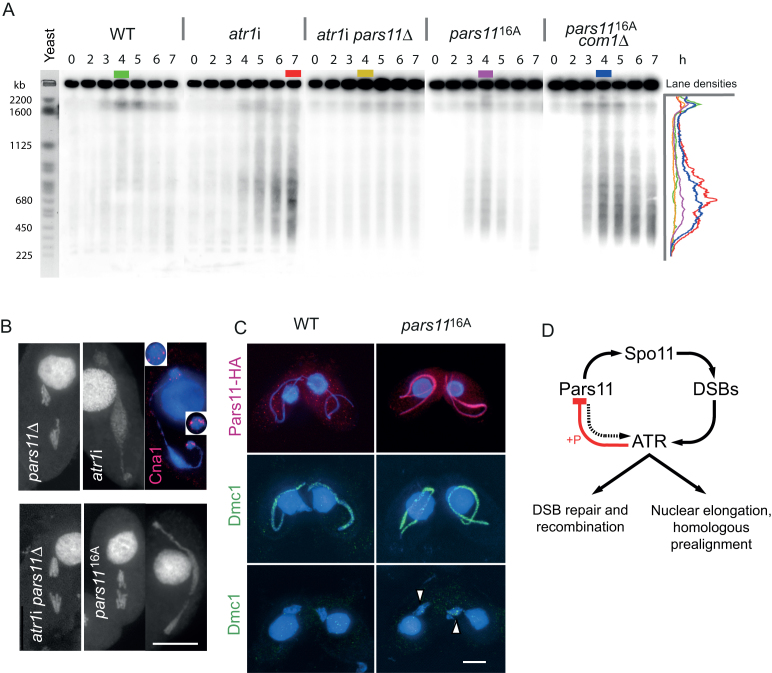
Figure 5.
Excessive DSB formation in the absence of ATR and phosphorylatable Pars11 and a model of ATR−Pars11 interaction. (A) In the WT, DSB-dependent fragments are transiently formed after onset of meiosis. In the absence of ATR, a few large and numerous smaller fragments indicate excessive DSB formation, which is suppressed by PARS11 deletion. In the presence of non-phosphorylatable Pars1116A, DSB formation is also increased. Increased DSB formation in pars1116A is particularly apparent when DSB repair is prevented in a double mutant with com1Δ. Density profiles (in arbitrary units) of lanes with the highest intensity (color-coded) show the size distribution of DSB-dependent fragments. Budding yeast chromosomes are shown as size markers. h: hours after induction of meiosis. (B) In pars11Δ, intact univalents separate in anaphase I. In atr1i, a chromatin mass assumes a dumbbell shape during the attempted anaphase. Cna1 staining indicates that kinetochore-containing chromosome parts migrate to opposing ends of the chromatin mass, whereas the bulk of chromatin does not separate (inserts show enlarged kinetochore regions). (Brightness of the somatic nucleus was selectively reduced to improve visualization of the adjacent germline nucleus.) In the atr1i pars11∆ double mutant, anaphase movement is restored. In the pars1116A mutant, effects on anaphase are variable (examples of a normal segregation and a bridge are shown). (C) HA-tagged Pars11 has disappeared by late prophase in the WT (see also Figure 4B), whereas HA-tagged Pars1116A persists to late prophase. Dmc1 staining is more intense in pars1116A than in the WT, and some Dmc1 foci remain in diakinesis-metaphase I nuclei (arrowheads). Foci in the somatic nucleus arise from antibody cross-reaction with Rad51. Bars: 10 μm. (D) Model of DSB control by Pars11 and ATR. Pars11 contributes to DSB-dependent nuclear elongation (dotted arrow). ATR downregulates its own activity via a feedback loop with Pars11 (red).figure at full with

