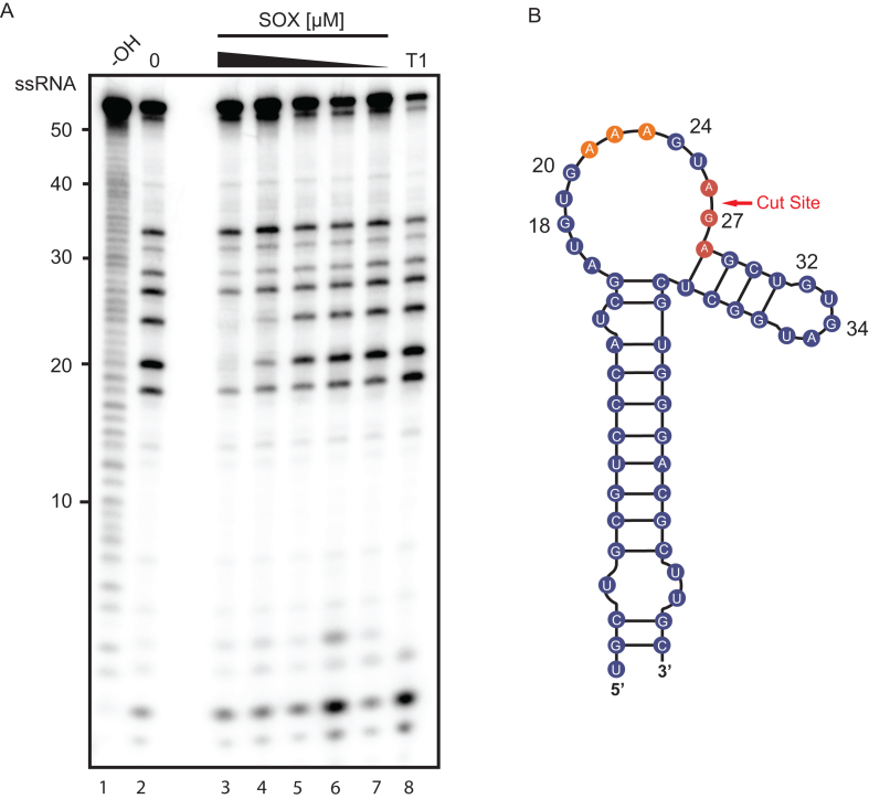
Figure 4.
SOX binds to a stretch of adenosines upstream of the cleavage site. (A) An RNA footprinting assay was carried out by incubating 5′ 32P-labeled LIMD1-54 with RNase T1 in the presence (lanes 3–7) or absence (lane 2) of a dilution series of SOX (8–0.5 μM). Hydrolysis (–OH, lane 1) and RNase T1 (T1, lane 8) ladders of the RNA were also generated in order to map the location of protected sites. Lines on the right denote protected base pairs. (B) Diagram of LIMD1-54 indicating sites protected from RNase T1 cleavage by SOX. The upstream SOX binding site is colored orange while the protected residues surrounding the cut site are shown in red.

