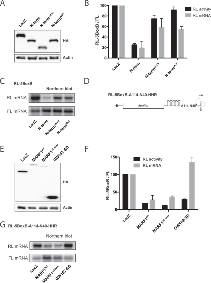
Figure 6.
MARF1 NYN displays endonuclease activity in vivo. (A and E) Expression levels of λNHA-tagged fusion proteins, as determined by Western analysis using anti-HA and anti-actin antibodies. (B) RL activity detected in extracts from HeLa cells expressing the indicated proteins. Cells were cotransfected with constructs expressing the RL-5BoxB reporter, FL, and indicated fusion proteins. Histograms represented normalized mean values of RL activity and mRNA levels from a minimum of three experiments. RL activity values seen in the presence of λNHA-LacZ were set as 100. (C) Representative Northern blot of RL-5BoxB and FL mRNAs levels for (B). (D) Schematic diagram of the Renilla luciferase-encoding mRNA reporter, containing five 19-nt BoxB hairpins, a 114 nt internal poly(A), a 40 nt linker and a self-cleaving hammerhead ribozyme (HHR). HHR cleavage site is denoted by an arrow. (F) Cells were cotransfected with constructs expressing the RL-5BoxB-A114-N40-HHR reporter, FL, and indicated fusion proteins. Histograms represented normalized mean values of RL activity and mRNA levels from a minimum of three experiments. RL activity values seen in the presence of λNHA-LacZ were set as 100. (G) Representative Northern blot of RL-5BoxB-A114-N40-HHR and FL mRNAs levels for (F).

