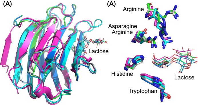
Figure 3. Comparison of three Gal-13 variant structures [R53HH57R (labeled by green), R53HH57RD33G (labeled by white) and R53HR33NH57RD33G (labeled by blue)] with Gal-3 CRD (labeled by cyan) and Gal-8 N CRD (labeled by purple).
(A) Global overlay. (B) The overlay of the sugar binding sites of Gal-13 variants, Gal-3 CRD, and Gal-8 N CRD. In Gal-13 variants, His53, Asn55, and Arg57 in Gal-13 variants show similar conformations to the corresponding residue of Gal-3 CRD and Gal-8 N CRD. Arg57 in R53HH57 and R53HH57RD33G shows two conformations. The galactose residues of lactoses in the sugar binding sites show similar conformations. However, the positions of galactose residues are not consistent.

