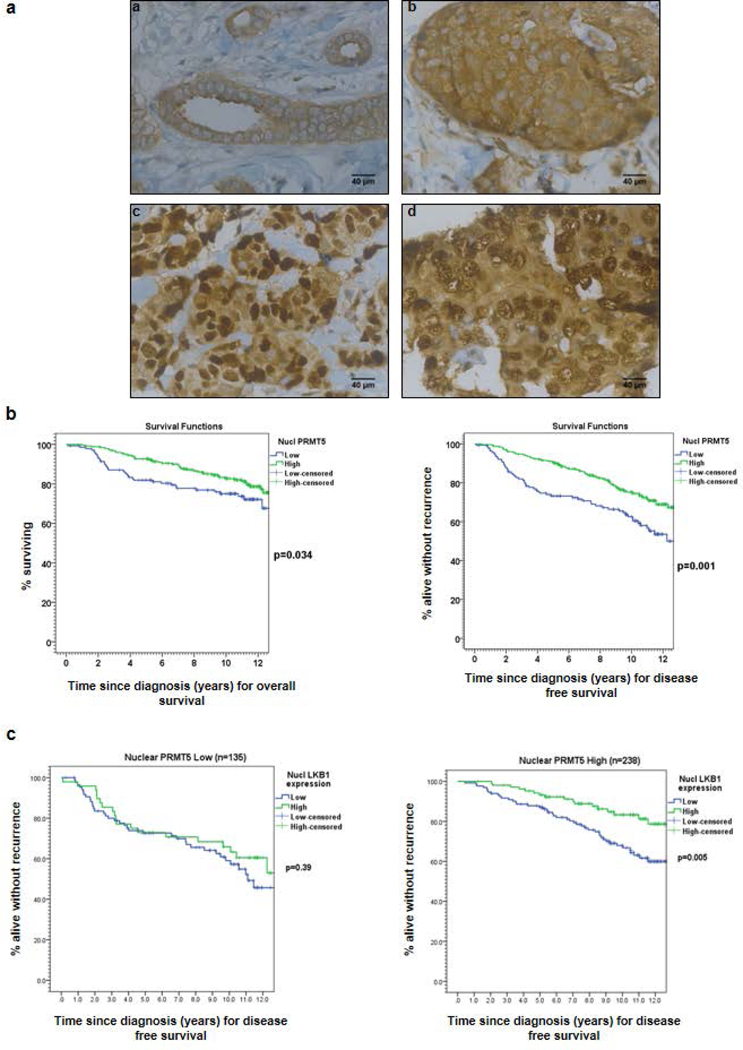
Figure 1: The expression of PRMT5 in breast tumors.

(a) PRMT5 expression was analyzed by IHC on formalin-fixed human tumors. Representative images of different IHC staining profiles are shown (panels a and b: cytoplasmic staining, panels c and d: cytoplasmic and nuclear staining) (Obj: X40). (b) Kaplan-Meier estimates of overall survival (OS) (left) and disease-free survival (DFS) (right) in patients with low (blue) versus high (green) nuclear PRMT5 expression. (c) Kaplan-Meier estimates of DFS in patients with low (blue) versus high (green) nuclear LKB1 expression in 2 groups of patients according to PRMT5 expression.
