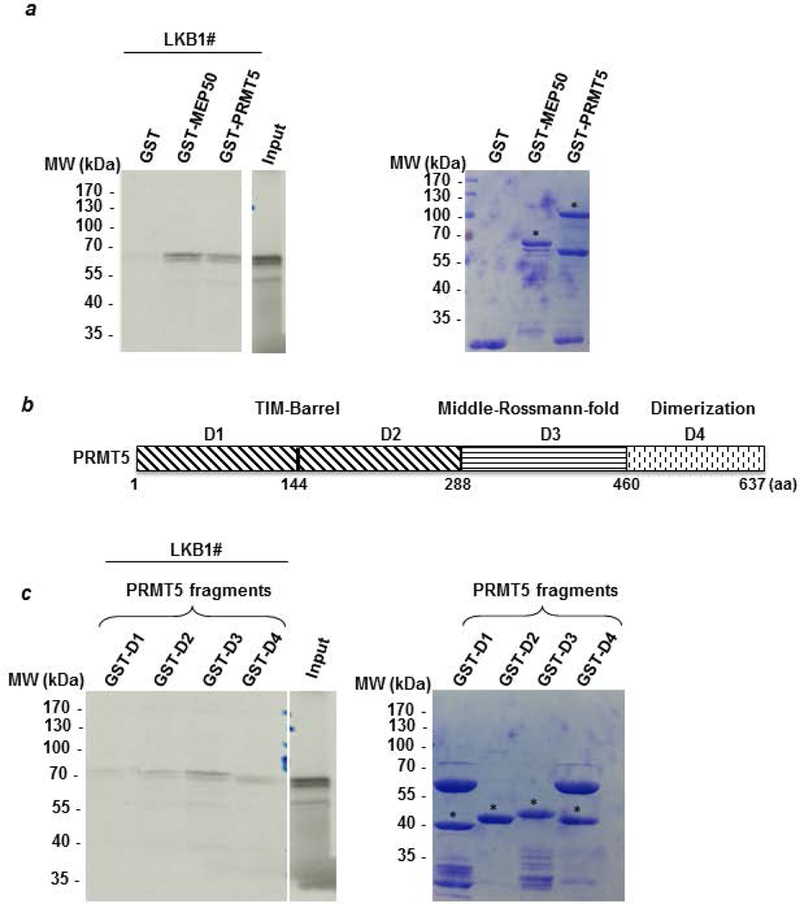
Figure 2: PRMT5 interacts with LKB1 in vitro.

(a) A radioactive GST pull-down assay was performed by incubating labeled in vitro 35S-labeled LKB1 (LKB1#) with GST, GST-MEP50 and GST-PRMT5. The corresponding Coomassie-stained gel is shown in the right panel. * indicates the full length fusion proteins. (b) PRMT5 was divided into 4 fragments (D1 to D4). The D1 and D2 fragments cover the TIM-Barrel domain. The D3 fragment corresponds to the Middle-Rossman-fold domain and the D4 fragment contains the dimerization domain. (c) Radioactive LKB1 (LKB1#) was incubated with the different domains of PRMT5 fused to GST and the bound proteins were visualized by autoradiography. The corresponding Coomassie-stained gel is shown in the right panel. # indicates the full length fusion proteins.
