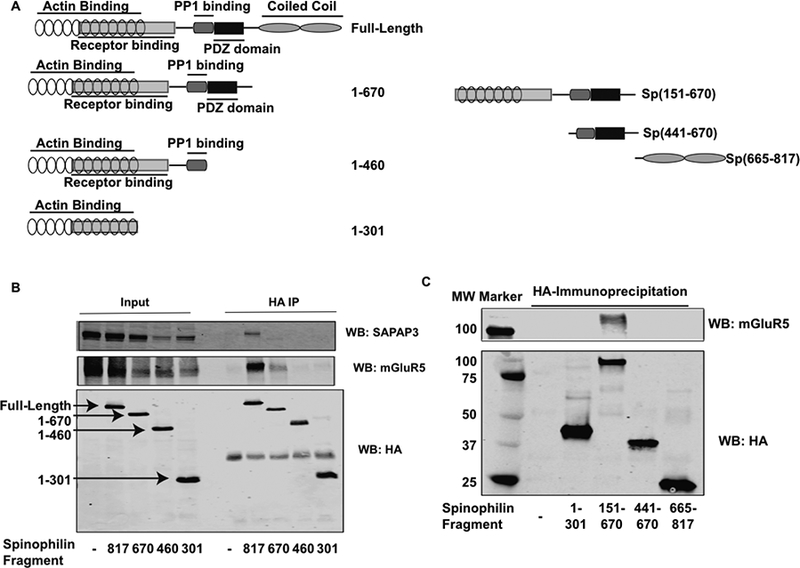
Figure 3. Mapping the domains on spinophilin that interact with SAPAP3 and mGluR5.

HEK293 cells were transfected with SAPAP3 and mGluR5 along with different deletion mutants of spinophilin. A. Schematic representation of spinophilin deletion mutants. Numbers refer to amino acids on different spinophilin fragments. B. HEK293 cells were transfected with SAPAP3 and mGluR5 alone or in the presence of different HA-tagged spinophilin deletion mutants. HEK293 cell lysates were immunoprecipitated with an HA antibody and immunoprecipitates were immunoblotted for SAPAP3, mGluR5, and HA. C. A different set of HA spinophilin fragments were immunoprecipitated and immunoblotted for HA and mGluR5. Experiments are representative of 3 independent trials.
