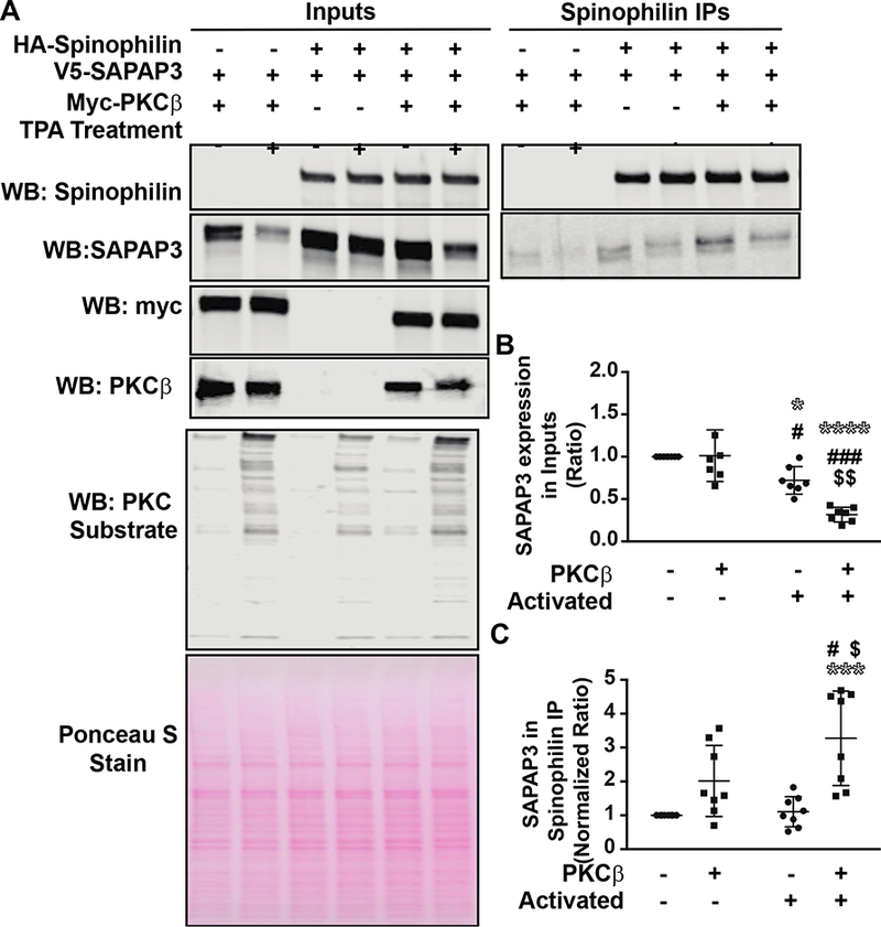
Figure 4. Activation of PKC𝛃 enhanced spinophilin/SAPAP3 interaction.

HEK293 cells were transfected with spinophilin and SAPAP3 in the absence or presence of PKCβ. The following day, HEK293 cells were treated with 200 nM TPA or vehicle for 30 minutes. A. Lysates were immunoprecipitated with an HA spinophilin antibody and lysates and immunoprecipitates were stained with Ponceau S and immunoblotted for spinophilin, SAPAP3, PKCβ, and PKC substrate substrate antibody. B. Activation of PKC and overexpression of PKCβ along with activation of PKC significantly decreased the expression of SAPAP3 compared to: no overexpression or activation (* symbol), PKCβ overexpression only (# symbol), or activation only ($ symbol). C. Activation of PKC along with overexpression of PKCβ significantly increased the association between spinophilin and SAPAP3. *,#,$ p<0.05, $$ p<0.01, ***, ###p <0.001, **** p<0.0001.
