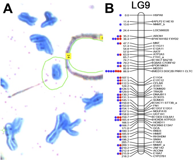
Figure 2.
Individual sex chromosome dyad alignment results on LG9. Read mapping was used to assess the specificity of the laser capture, amplified library of the sex chromosome dyad. (A) A partial metaphase spread of axolotl chromosomes stained with Giemsa on a membrane slide. The sex chromosome is circled in green. (B) The distribution of markers sampled from the sex chromosome (LG9) via targeted sequencing of individual chromosomes. LG9 is based on a previously published linkage map for the axolotl35. Individual gene markers are designated by labels to the right of their corresponding map position and their predicted position (in centiMorgans) is provided by numerical labels to the left. Dots represent markers with mapped reads from a single library. Red denotes the first sequencing attempt using the DNA-seq kit with 48 total barcoded samples on a single lane of an Illumina HiSeq flowcell. Blue denotes re-sequencing of the same chromosome library on a single lane.

