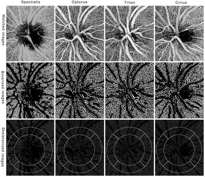
Figure 3.
OCTA en face images of four devices. After registration, the four original images of four devices display exactly the same location (upper row). After a process of Hessian filter, auto-threshold and large vessel extraction, we got the binarized images (middle row). The images show single lines of retinal vessels after skeletonization and the selected locations for measurement are overlaid on the skeletonized images (bottom row). The diameters of inner and outer circles are 1.5 mm and 2.25 mm, respectively.

