Fig. 4.
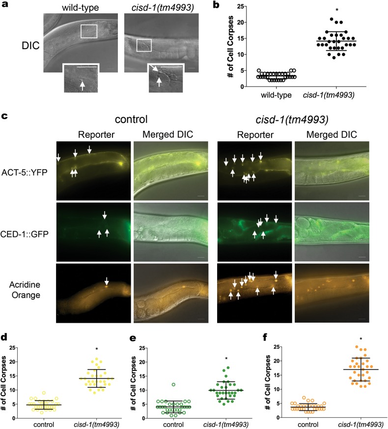
The cisd-1(tm4993) animals exhibits an increase in germline cell corpses. a Representative DIC images of one gonad arm with cells undergoing apoptosis in the cisd-1(tm4993) and N2 wild-type animal. The boxed region shows an enlarged area containing the cell corpse(s); the white arrow points to an apoptotic cell appearing as a “button”. Scale bar = 20 µm. b The cisd-1(tm4993) animal has a significantly higher number of apoptotic cells relative to the N2 wild-type as determined by analysis of the gonad using DIC microscopy. c Representative fluorescent microscopy images of the germline for control or cisd-1(tm4993) animals. Reporters were used to mark specific aspects of programmed cell death (ACT-5::YFP, CED-1::GFP, or acridine orange). Shown are the fluorescent reporters individually or merged with the DIC image to better visualize the gonad anatomy. White arrows mark either germ cell corpses with surrounding actin bundles in the early stages of apoptosis (ACT-5::YFP), surrounding cells that have initiated engulfment (CED-1::GFP), or cells positive for cell surface acridine orange staining. Scale bar = 20 µm. d-f Relative to control populations, the cisd-1(tm4993) animals have a significantly higher number of germline apoptotic cells marked by d ACT-5::YFP, e CED-1::GFP positive cells, or f acridine orange. For b, d, e and f, the number of apoptotic corpses within the gonad was quantified in animals from three independent experiments for a total of at least 30 animals (*indicates P < 0.0001, two-tailed unpaired parametric t-test, or Mann–Whitney two-tailed nonparametric test). The error bar indicates standard deviation.

