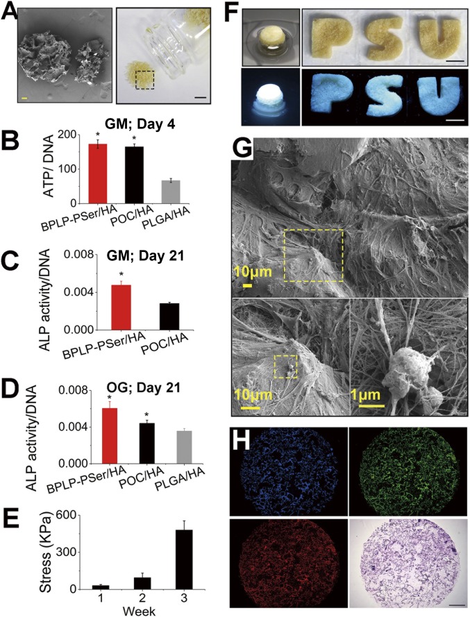
Fig. 5.
BPLP-PSer/HA MP scaffolds promote hMSC differentiation. (A) Photographic (Right) and SEM (Left) images of BPLP-PSer/HA MP scaffolds. (Scale bars: Right, 5 mm; Left, 100 µm.) (B) Intracellular ATP levels (normalized to DNA) of hMSCs cultured on different MPs in GM for 4 d. n ≥ 8 biological replicates per group. (C and D) ALP production of hMSCs in GM without osteogenic inducers cultured on MPs in Transwell 3D models (C) and differentiated in OG medium for 21 d (D). n = 3–5 biological replicates per group. (E) Compressive strength of round disk-shaped cell–MP constructs after cells differentiated for 1, 2, or 3 wk in OG medium. n = 3 cell–MP constructs per time point. All data are presented as mean ± SD; *P < 0.05. (F) Photographic and fluorescent images of hMSC–MP constructs obtained with 21 d of culture in round Transwells (Left) or from in PDMS wells with permeable bottoms cast from 3D-printed letter molds in the shape of the letters P, S, and U (Right). (Scale bars: 5 mm.) (G) SEM images of the thick cell layer covering and bridging MPs (Upper) and the extensive interwoven ECM network (Lower Left) produced by hMSCs differentiated for 21 d in OG medium to enable mineral formation (Lower Right). (H) Fluorescent images (blue, green, and red channels) (Upper Left, Upper Right, and Lower Left, respectively) and H&E staining (Lower Right) of the cell–MP construct sections obtained by cryo-sectioning. (Scale bar: 1 mm.)

