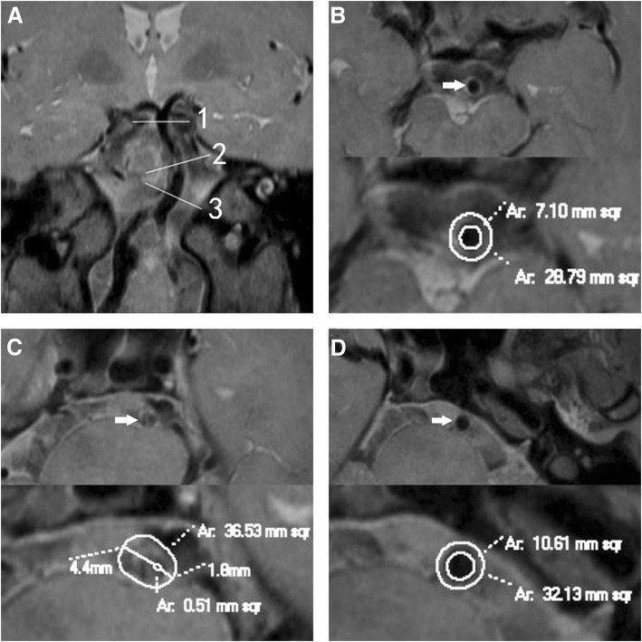
Fig. 1.
Positive remodeling. A patient (61–65 years) with hypertension and smoking history presented with dizziness for 2 months. Coronal reconstruction image a revealed the plaque in BA distal to AICA. The line 1 represents the distal reference site; line 2, maximal-lumen-narrowing (MLN) site; line 3, proximal reference site. Cross-sectional images at the distal, MLN and proximal sites were shown in Figs. b, c and d respectively, as shown by the arrow. The vessel area (VA) is 36.53 mm2 at MLN site, 28.79 mm2 at distal site, and 32.13 mm2 at proximal site. The reference VA is 30.46 mm2. The remodeling index (RI) is 1.20 (RI ≥ 1.05, defined as positive remodeling). The lumen area (LA) is 0.51 mm2 at MLN site, 7.10 mm2 at distal site, and 10.61 mm2 at proximal site. The reference LA is 8.86 mm2. The wall area (WA) is 36.02 mm2 at MLN site, and 21.6mm2 at reference site. So the plaque size is 14.42 mm2. The maximal wall thickness is 4.4 mm, and the minimal wall thickness is 1.8 mm. The eccentricity index is 0.59, and percentage of plaque burden is 39.5%. The plaque was distributed eccentrically and predominantly located at the right wall

