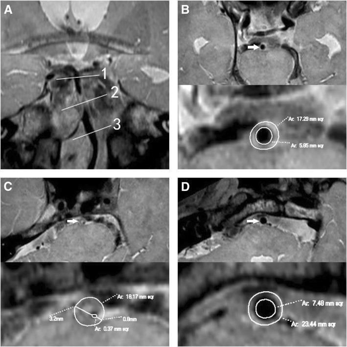
Fig. 2.
Negative remodeling. A patient (56–60 years) with hypertension, diabetes mellitus and hyperlipidemia presented with left limb weakness for 4 days. Coronal reconstruction image a revealed the plaque in BA distal to AICA. The line 1 represents the distal reference site; line 2, maximal-lumen-narrowing (MLN) site; line 3, proximal reference site. Cross-sectional images at the distal, MLN and proximal sites were shown in Figs. b, c and d respectively, as shown by the arrow. The plaque was located in BA distal to AICA. The VA is 18.17 mm2 at MLN site, 17.29 mm2 at distal site, and 23.44 mm2 at proximal site. The reference VA is 20.37 mm2. The remodeling index (RI) at the MLN site was 0.89 (RI ≤ 0.95, defined as negative remodeling). The LA is 0.37 mm2 at MLN site, 5.85 mm2 at distal site, and 7.48 mm2 at proximal site. The reference LA is 6.67 mm2. The WA is 17.8 mm2 at MLN site, and 13.7 mm2 at reference site. So the plaque size is 4.1 mm2. The maximal wall thickness is 3.2 mm, and the minimal wall thickness is 0.8 mm. The eccentricity index is 0.75, and percentage of plaque burden is 22.6%. Compared with the case above (Fig. 1), this one had smaller VA, WA, plaque size and percentage of plaque burden

