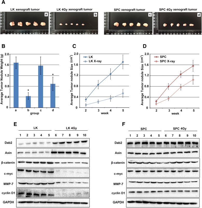
Fig. 6.
The effect of X-ray irradiation on xenograft tumor growth in LK and SPC cells. a Photograph of mice and excisional xenograft tumors inoculated with LK cells (a), 4Gy irradiation treated LK cells (b), SPC cells (c) and 4Gy irradiation treated SPC cells (d) for 5 weeks. More significant inhibition was observed in LK irradiated cells rather than SPC irradiated cells (P < 0.05). b Histogram representation of average tumor weight of each group. X-ray irradiation was more effective in LK cells rather than SPC cells (P < 0.05). c and d. Average tumor volumes of LK and SPC cells after irradiation, respectively. The results showed more significant inhibition in LK cells at 2, 3, 4 and 5 week than SPC cells (P < 0.05). e Dab2 and Axin were up-regulated in LK xenograft tumors with X-ray treatment comparing with un-treated group (1–5 VS 6–10 P < 0.05), and decreased expression of β-catenin, c-myc, MMP-7 and cyclinD1 were also identified in irradiation group but not in un-treated group (1–5 VS 6–10 P < 0.05). f No obvious difference of Dab2 and other factors of Wnt pathway were identified between SPC xenograft tumors with X-ray treatment and un-treated group (1–5 VS 6–10). * P < 0.05

