Figure 2.
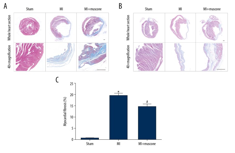
Muscone reduced fibrosis in left ventricular (LV) myocardium after MI for 4 weeks. Representative histological photomicrographs showed the collagen deposition (blue) on the infarct region in transverse sections (A) and longitudinal sections (B) in each group as shown by Masson’s trichrome staining. Scale bars=500 μm. (C) Quantitative analysis of fibrotic area by Masson’s trichrome staining. Data are represented as mean ±SD, n=4 per group (* p<0.05 versus sham group, # p<0.05 versus MI group).

