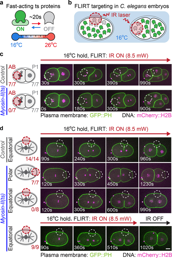
Figure 1:
FLIRT calibration and application for spatiotemporal control of ts protein function in vivo
(a) Fast-acting ts mutant proteins are rapidly inactivated upon temperature upshift3, 6. (b) Schematic of FLIRT targeting in which an IR laser is used to locally inactivate ts mutant protein function. (c) Experimental schematic (left) and representative time lapse images (right) of cell-specific FLIRT targeting in 2-cell C. elegans embryos. See Supplementary Video 1. (d) Experimental schematic (left) and representative time lapse images (right) of subcellular FLIRT targeting either an equatorial or polar region in myosin-II(ts) 1-cell embryos. Embryos were FLIRT-targeted either throughout division (top 3 rows, see Supplementary Video 3) or for ~8 min to test reversibility (bottom row, see Supplementary Video 5). The number of AB and P1 cells (c) or 1-cell embryos (d) from biologically independent embryos that successfully completed cell division is indicated below each experimental schematic (left). Red (schematic) or white (images) dashed circles indicate the FLIRT-targeted regions. Time is in sec after FLIRT initiation. Scale bars=10 μm.

