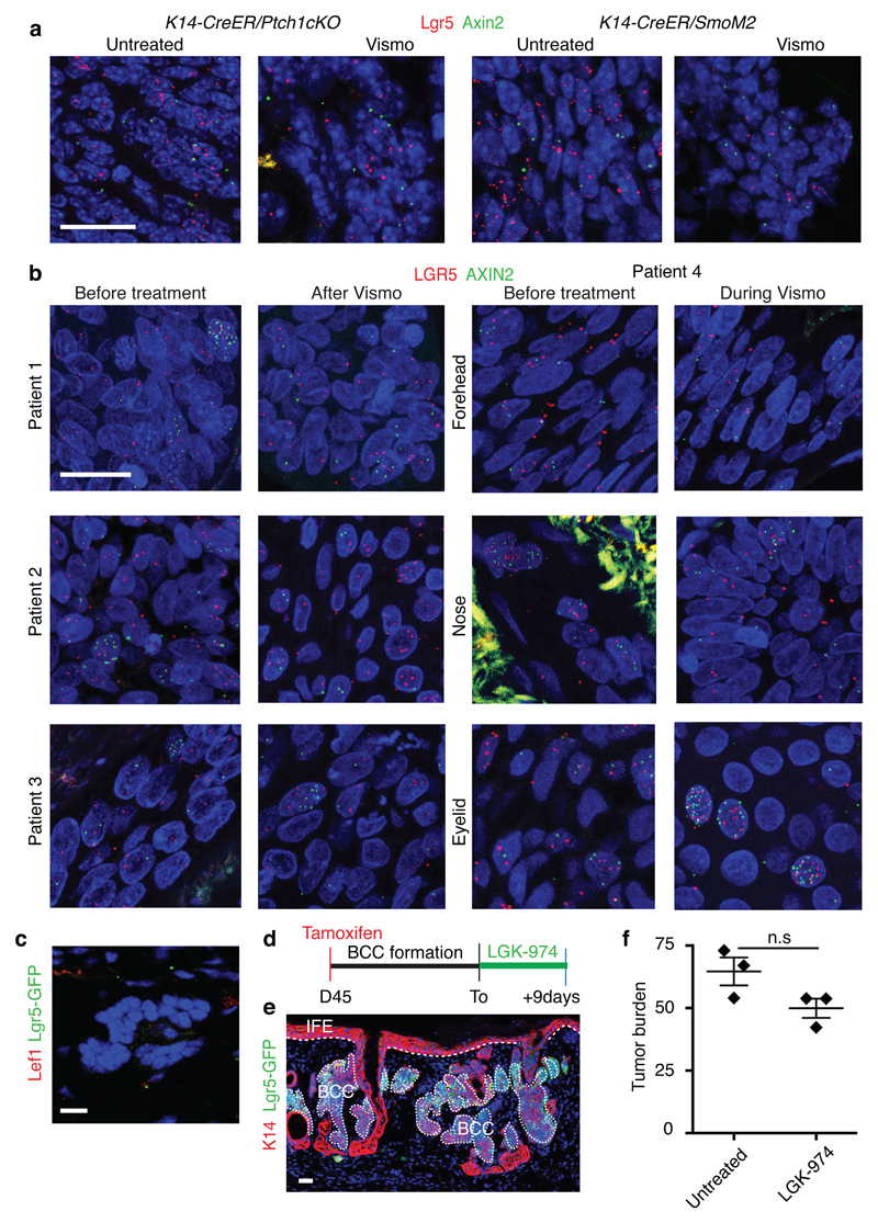
Extended Data Fig. 10. Wnt signalling is active in mouse and human vismodegib-persistent lesions.
(a) In situ hybridization for Lgr5 and Axin2 in untreated and vismodegib-treated lesions from Ptch1cKO and SmoM2 mice.(b) In situ hybridization for LGR5 and AXIN2 in biopsies from patients before, during and after vismodegib treatment. (c) Immunostaining for Lef1 and GFP in Ptch1cKO/Lgr5-DTR-FP-derived tumorigenic lesion following vismodegib+ LGK-974 treatment. (d) Protocol used for LGK-974 treatment in Ptch1cKO/Lgr5-DTR-GFP mice (e) Immunostaining for GFP and K14 in BCC treated with LGK-974 for 9 days from the Ptch1cKO/Lgr5-DTR-GFP model. (f) Quantification of the tumour burden in BCC treated with LGK-974 for 9 days and untreated (n=3 Ptch1cKO/Lgr5-DTR-GFP mice). Description of the skin length and tumour area analysed per mouse in Source Data. Two-sided t-test. Mean+/- s.e.m. Three independent experiments per condition were analysed showing similar results (a,e) and two technical replicates were performed for each sample showing similar results (b).Hoechst nuclear staining in blue; scale bars, 25 μm. IFE: interfollicular epidermis, BCC: basal cell carcinoma, HF: hair follicle. Dashed line delineates basal lamina separating IFE from the dermis. Dotted line delineates BCC.

