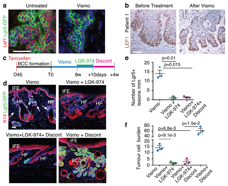
Fig. 4. Dual HH and Wnt inhibition eliminates vismodegib-persistent Lgr5+ TCs.
(a) Immunostaining for GFP and Lef1 in Ptch1cKO/Lgr5-DTR-GFP mice untreated and vismodegib-treated. (b) Immunohistochemistry for LEF1, in biopsies from a patient before and after vismodegib treatment. (c) Protocol for dual HH and Wnt inhibition followed by treatment discontinuation. (d) Immunostaining for GFP and K14 upon vismodegib administration, dual inhibition of Wnt and HH pathways and following discontinuation in Ptch1cKO/Lgr5-DTR-GFP mice. (e) Number of Lgr5+ tumorigenic lesions per length of epidermis upon treatment and treatment discontinuation in Ptch1cKO/Lgr5-DTR-GFP-derived BCCs. (n=3 mice, 3mm of skin analysed per mouse). Mean+/- s.e.m. Two-sided t-test. (f) Quantification of the tumour burden upon treatment and treatment discontinuation in Ptch1cKO/Lgr5-DTR-GFP-derived BCCs (n=3 mice). See Source Data. Two-sided t-test. Mean+/- s.e.m. Three independent experiments per condition were analysed showing similar results (a) and two technical replicates were performed for each sample showing similar results (b). Hoechst nuclear staining in blue; scale bars, 50 μm. Dashed line delineates basal lamina Arrow indicates vismodegib-persistent lesions.

