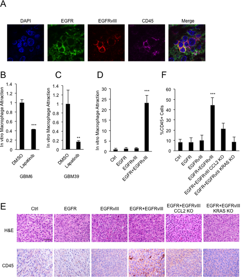
Figure 1.

Co-expression of EGFR and EGFRvIII leads to infiltration of CD45+ cells. A, Cells double positive for EGFR and EGFRvIII colocalize with CD45+ cells in sections from primary EGFR/EGFRvIII+ human glioblastoma. B and C, Transwell migration assay showing attraction of macrophages (macrophage line: MV-4–11) in response to conditioned media from GBM6 and GBM39 cells treated with DMSO or 5μM lapatinib for 24 hours. D, Transwell migration assay showing attraction of macrophages (macrophage line: MV-4–11) in response to conditioned media from U87 cells co-expressing EGFR and EGFRvIII; E, H&E and IHC staining validating increased numbers of CD45 positive cells in intracranial xenografts driven by EGFR and EGFRvIII; and decreased numbers of CD45 positive cells in xenografts deleted for CCL2 and KRAS; F, quantification. Macrophage attraction levels were normalized to the control cell line. Results were reproduced in three independent experiments with triplicate samples. **, p<0.01, ***, p < 0.001. Scale bar: 50μM.
