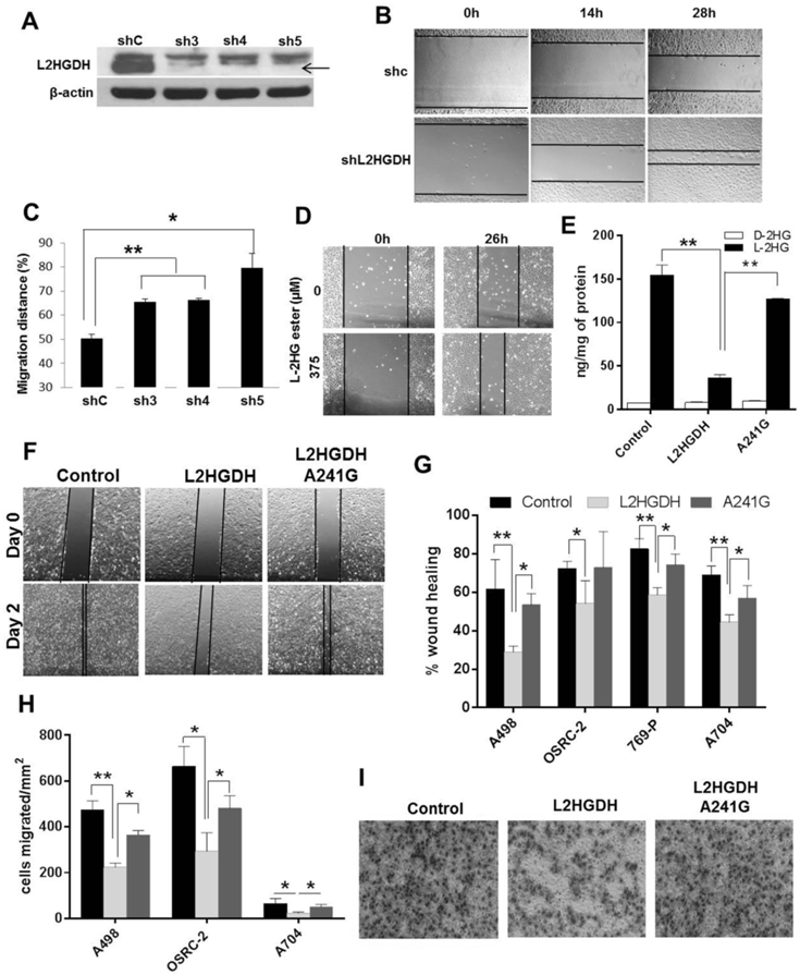
Figure 1. The L2HGDH/L-2-HG axis regulates migratory phenotypes.

(A) HK-2 cells were transduced with control shRNA or three shRNAs targeting L2HGDH (sh3, sh4 and sh5) and puromycin resistant cells were selected to generate pooled stable cell lines. Validation of L2HGDH knockdown by immunoblotting (black arrow). (B and C) Wound healing of shL2HGDH stable cell lines seen using time-lapse phase contrast photography. Migration distance (%) calculated at 28 hr post-wound healing. Data are representative of two independent experiments (n=3/group). (D) Representative bright field images captured 26 hr post-wound creation in HK-2 cells treated with L-2-HG octyl ester. Data are representative of two independent experiments. (E) LC-MS analysis of L-2-HG and D-2-HG levels in control vector, WT L2HGDH, A241G expressing A498 cells. (F) Representative bright field images of A498 cells at day 0 and day 2 post-wound creation. (G) Relative wound healing of A498, OSRC-2, 769-P, A704 stably expressing control, L2HGDH and A241G vectors at 48, 36, 24 and 120 hrs post-wound creation, respectively. Data shown are the means ± SEM of two independent experiments (n=3/group). (H) Quantification of RCC cell migration (A498, OSRC-2, and A704) expressing control, L2HGDH and A241G. Data shown are the means ± SEM of two independent experiments (n=3/group). (I) Representative images of cell migration of A498 cells stably expressing control, L2HGDH and A241G vectors after 16 hrs incubation. (* indicates p < 0.05, ** indicates p < 0.01).
