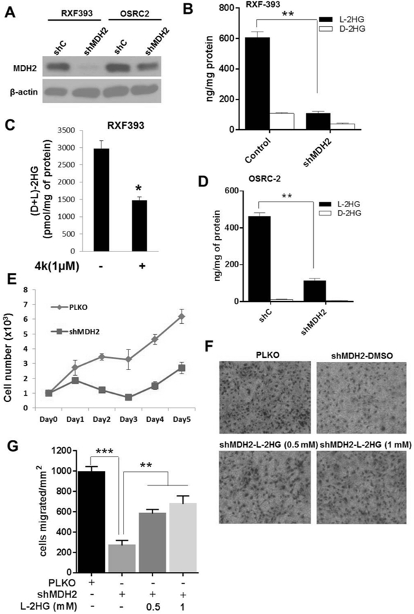
Figure 4. Knockdown of MDH lowers L-2-HG and suppresses in vitro tumor phenotypes in RCC cells.

OSRC-2 and RXF-393 cells were transduced with PLKO control and shMDH2 vectors. (A) Western blot analysis of MDH2 knockdown in RXF-393 and OSRC-2 cells. (B) Intracellular L-2-HG and D-2-HG level in shMDH2 transduced RXF-393 cells. (C) RXF-393 cells were treated with MDH inhibitor (4k, 1uM) for 48 h, harvested, and assayed for total 2-HG levels. (D) Intracellular L-2-HG and D-2-HG levels in PLKO and shMDH2 transduced OSRC-2 cells. (E) Proliferation of OSRC-2 cells transduced with control PLKO and shMDH2 vector. Data shown are the means ± SEM of two independent experiments (n=3/group). (F, G) OSRC-2 cells transduced with shMDH2 were treated with or without L-2-HG ester (0.5 and 1 mM) for 48 hrs and allowed to migrate in Boyden’s chamber for 16 hrs. (F) Representative images of OSRC-2 cells migrated in Boyden’s chamber. (G) Quantification of OSRC-2 cells migrated in Boyden’s chamber. Data shown are the means ± SEM of two independent experiments (n=3/group). (* indicates p < 0.05, ** indicates p < 0.01, *** indicates p < 0.001).
