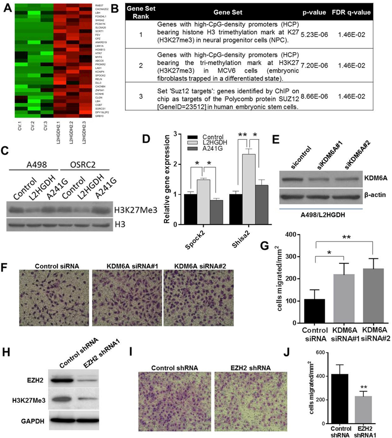
Figure 5. High L-2-HG inhibits activity of histone lysine demethylase to promote H3K27 trimethylation in RCC cells.

(A) Heat map of PRC2/H3K27me3 target genes with increased expression upon L2HGDH restoration (n=3/group). (B) GSEA of genes with increased expression upon L2HGDH restoration. (C) Immunoblots of H3K27Me3 levels in A498 and OSRC-2 cells expressing control, WT L2HGDH, and L2HGDH A241G. (D) Relative mRNA levels of SPOCK2 and SHISA2 in A498 cells stably expressing control, WT L2HGDH, and L2HGDH mutant A241G measured using RT-qPCR. (E) A498 cells expressing L2HGDH were treated with the indicated siRNA and then assessed by immunoblotting for KDM6A protein levels. (F) Representative images of A498/L2HGDH cells treated with the indicated siRNA migrated through a transwell insert. (G) Quantification of migration of A498/L2HGDH cells treated with the indicated siRNA. Data shown are the means ± SEM of two independent experiments (n=3/group). (H) Immunoblot for EZH2 and H3K27me3 in A498 cells transduced with control or EZH2 shRNA. (I) Representative images of A498 cells transduced with the indicated shRNA migrated through a transwell insert. (J) Quantification of migration of A498 cells transduced with the indicated shRNA. Data shown are the means ± SEM of two independent experiments (n=3/group). (* indicates p < 0.05, ** indicates p < 0.01).
