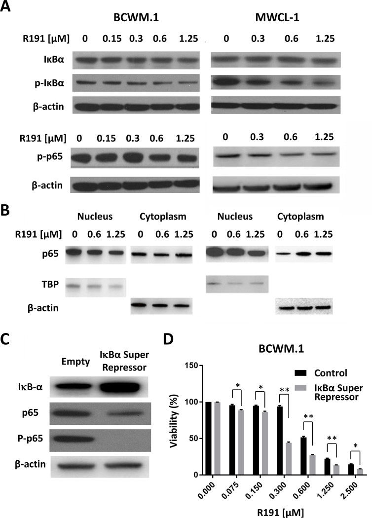
Figure 3. Inhibition of NF-κB in R191-treated cell lines.

BCWM.1 and MWCL-1 cells were treated with indicated R191 concentrations for 24 hours. The NF-κB activation status was evaluated by Western blotting of whole cell lysates to determine the expression and phosphorylation status of IκBα and the p65 subunit of NF-κB (A), or of nuclear and cytoplasmic fractions for the p65 subunit (B). β-actin was used as the control for total lysates and cytoplasmic fractions, while TBP was used for the nuclear fraction. Also, BCWM.1 cells expressing an IκBα super-repressor (S32A/S36A) or vector control sequences were generated (C) in which the super-repressor reduced phospho-p65 levels. These cells were then exposed to the indicated R191 concentrations for 24 hours (D), and cell viability was determined. Data are expressed as the means ± SD for samples assayed in triplicate, and “*” indicates p-values <0.003, while “**” indicates p-values <0.0001 for the noted comparisons.
