
Figure 3:
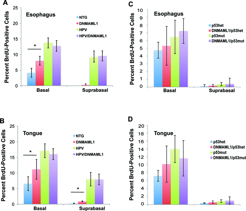
Analysis of cellular proliferation with loss of Notch signaling on an HPV-positive or HPV-negative background. To determine basal and suprabasal cellular proliferation in the tongue and esophagus, mice were injected intraperitoneally with BrdU prior to sacrifice. The tissue was then stained via immunohistochemistry against anti-BrdU. A minimum of 3 mice from each genotype (NTG, DNMAML1/K14CreER, E6/E7, DNMAML1/K14CreER/E6/E7) were stained, and 8 to 10 sections per mouse were captured at 200 times magnification. Each section was scored for percentage of basal and suprabasal proliferation using ImageJ. A) Average Proliferation for the esophagus for the HPV-positive cohort. p-value via Student’s T-Test for basal NTG versus DNMAML1 = 0.009. B) Average proliferation for the tongue for the HPV-positive cohort. p-value via Student’s T-Test for basal NTG versus DNMAML1 = 0.042, for suprabasal NTG versus DNMAML1 < 0.001. C) Average proliferation for the esophagus for the HPV-negative cohort. D) Average proliferation for the tongue for the HPV-negative cohort. p53mut mice exhibited slightly higher proliferation in the tongue and esophagus with or without DNMAML1, but this difference was not significant. DNMAML1 did not increase cellular proliferation in the tongue or esophagus.