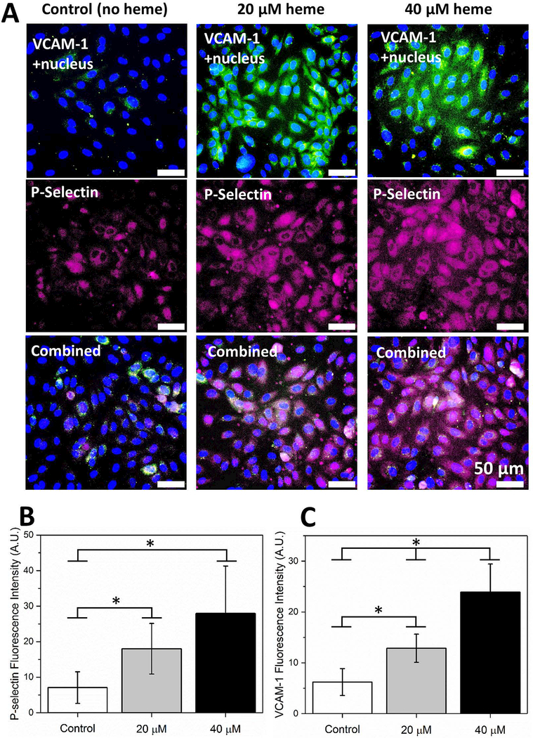
Figure 2. Activation of ECs with heme results in VCAM-1 and P-selectin expression in a concentration dependent manner.
(A) ECs were treated with RPMI containing 0 μM, 20 μM, and 40 μM heme for 60 minutes in 37 °C and incubated with fluorescently labeled antibodies against VCAM-1 and P-selectin following a fixing step with 4% PFA. Cell nuclei were stained with DAPI. (B) Expression of P-selectin was significantly elevated in 20 μM and 40 μM heme-treated ECs (p<0.05, one-way ANOVA) compared to quiescent ECs while a significant difference was absent between 20 μM and 40 μM heme-treated ECs. (C) Similarly, VCAM-1 expression was significantly greater in heme-activated ECs in comparison with quiescent ECs, 40 >20 μM heme (P<0.05). The horizontal brackets and stars between different groups indicate statistically significant difference based on a one-way ANOVA test (n=5 in each group, P<0.05). Error bars = 50 μm.

